
Dr. James Manos (MD)
January 5, 2016
Review: Tips in Medical Biochemistry
Volume (4)
CONTENTS
OTHER METABOLIC DISTURBANCES
Glucose
Insulin
Hyperglycemia
Glycemic index (GI) & glycemic load (GL)
Glycemic index (GI) & glycemic load (GL) calculator
Hypoglycemia
Pseudohypoglycemia (artifactual hypoglycemia) – causes
Diagnostic algorithm for hypoglycemia
Uric acid
Gout & uric acid levels
Uric acid in diet and recommendations for people with gout
OTHER METABOLIC DISTURBANCES
Glucose
· Insulin: is an anabolic hormone that promotes glucose uptake, glycogenesis, lipogenesis, and protein synthesis of skeletal muscle and fat tissue through the tyrosine kinase receptor pathway. Also, it is the most crucial factor in the regulation of plasma glucose homeostasis (regulation), as it counteracts glucagon and other catabolic hormones—epinephrine, glucocorticoid, and growth hormone. A standard insulin test is positive for endogenous insulin and exogenous insulin. Also, there is a minimal cross-reaction with proinsulin and insulin-like growth factors 1 and 2, with the degree of variability depending on the brand of the testing toolkit and technique used.
· Interpretation: Insulin testing is used to assist in identifying causes of hypoglycemia (plasma glucose levels < 55 mg/dL), especially upon signs and symptoms of hypoglycemia (neuro-hypoglycemic and autonomic symptoms). In this case, a 72-hour fasting test is performed.
· On insulinoma (a tumor of the pancreas that is derived from beta cells and secretes insulin) we have high insulin and C-peptide levels.
· On non–beta-cell tumors we have low insulin and C-peptide levels and high insulin-like growth factor 2 levels.
· On excessive insulin administration, we have high insulin levels and low C-peptide levels.
· On insulin secretagogue administration (sulfonylurea and glinides; both are antidiabetic medications) we have high insulin and C-peptide levels.
· On congenital hyperinsulinism (mutation in an insulin-secreting gene) we have high insulin and C-peptide levels.
· On autoimmunity to insulin or insulin receptor (common in patients receiving insulin or those who have autoimmune diseases such as systemic lupus erythematosus or Hashimoto thyroiditis): postprandial (after a meal) insulin is bound to antibodies and dissociated 1 hour later, resulting in an extremely elevated insulin level and high insulin–to–C-peptide ratio.
· Causes of elevated insulin levels:
· a) Conditions associated with increased insulin resistance (beta-cell compensates via hypersecretion of insulin) include obesity, steroid administration, acromegaly, Cushing syndrome, insulin receptor mutation, and type 2 diabetes (early stage).
· b) Conditions associated with increased insulin secretion include insulinoma (insulin or proinsulin-secreting tumors) and administration of insulin secretagogues (such as sulfonylurea and glinides antidiabetic medications). Excessive administration of insulin is associated with elevated insulin levels.
· c) Conditions associated with decreased insulin excretion include severe liver disease and severe heart failure (liver congestion).
· Autoimmunity to insulin or insulin receptor is associated with elevated insulin levels.
· Causes of decreased insulin levels: conditions related to beta-cell destruction include: post (after) pancreatectomy; chronic pancreatitis; autoimmune destruction; type 1 diabetes.
· In type 2 diabetes (late stage), beta cells fail to secrete insulin to maintain the blood glucose level, owing to insulin resistance and genetic defect.
· Increased insulin-like growth factor (IGF) levels are associated with non–beta-cell tumors.
· A 72-hour fasting test is used to identify the causes of postabsorptive hypoglycemia. The patient is instructed to fast, and plasma glucose, insulin, proinsulin, and C-peptide levels are measured every 6 hours until the plasma glucose level is less than 65 mg/dL, after which the testing frequency is increased to every 1 – 2 hours. Fasting is ended when plasma glucose levels are less than 45 mg/dL accompanied by signs and symptoms of hypoglycemia. A blood sample is collected and tested at the endpoint for glucose, insulin, proinsulin, C-peptide, beta-hydroxybutyrate, and sulfonylurea levels. The patient is given 1 mg of intravenous glucagon, and the response of the blood glucose level is measured.
· Interpretation of 72-hour fasting test:
· a) Insulinoma: increased insulin; increased C peptide; increased pro-insulin; decreased insulin growth factor 2 (IGF – 2); reduced sulfonylurea levels; increase of glucose level after administration of glucagon.
· b) Non–beta–cell tumors: decreased insulin; decreased C peptide; decreased proinsulin; increased insulin growth factor 2 (IGF – 2); reduced sulfonylurea levels; increased glucose level after administration of glucagon.
· c) Insulin injection: increased insulin; decreased C peptide; decreased proinsulin; decreased insulin growth factor 2 (IGF – 2); decreased sulfonylurea levels; increase of glucose level after administration of glucagon.
· d) On sulfonylurea (an antidiabetic medication) – induced: increased insulin; increased C peptide; increased pro-insulin; decreased insulin growth factor 2 (IGF – 2); increased sulfonylurea levels; increase of glucose level after administration of glucagon.
· Hyperglycemia: hyperglycemia or high blood sugar is a condition in which an excessive amount of glucose circulates in the blood plasma. This is generally a blood sugar > 11.1 mmol/l (200 mg/dl), but symptoms may not start to become noticeable until even higher values, such as 15–20 mmol/l (about 250 – 300 mg/dl). A subject with a consistent range between about 5.6 and 7 mmol/l (100–126 mg/dl) according to the American Diabetes Association guidelines, is considered hyperglycemic, while above 7 mmol/l (126 mg/dl) is generally held to have diabetes mellitus. Chronic levels exceeding 7 mmol/l (125 mg/dl) can produce organ damage.
· High glucose levels most frequently indicate diabetes, but many other diseases (described below) and conditions can also cause elevated blood glucose. In a person with signs and symptoms of diabetes or hyperglycemia, a non-fasting glucose level (random blood sample) equal to or greater than 200 mg/dL (11.1 mmol/L) indicates diabetes. A blood glucose test may also be ordered when someone has signs and symptoms of high blood glucose.
· Signs & symptoms: polydipsia (increased thirst), usually with polyuria; fatigue; blurred vision; slow-healing wounds or infections; weight loss; dry mouth (xerostomia); fry or itchy skin; tingling in feet or heels; erectile dysfunction; recurrent infections (e.g., swimmer’s ear), etc.
· Diabetes screening: several health organizations, including the American Diabetes Association (ADA) and the U. S. Preventive Services Task Force (USPSTF), recommend diabetes screening when a person is age 45 or older or when a person of any age has risk factors.
· Examples of risk factors include: overweight, obese, or physically inactive; a close (first degree) relative with diabetes; a woman who delivered a baby weighing more than 9 pounds (about 4 kg) or with a history of gestational diabetes; a woman with the polycystic ovarian syndrome (PCOS); high-risk race or ethnicity such as African American, Latino, Native American, Asian American, Pacific Islander; high blood pressure or taking medication for high blood pressure; low HDL (also known as ‘good’ cholesterol) level (less than 35 mg/dL or 0.90 mmol/L) and/or a high triglyceride level (more than 250 mg/dL or 2.82 mmol/L); A1c equal to or above 5.7%; prediabetes identified by the previous testing; and history of cardiovascular disease (CVD).
· Causes of hyperglycemia:
· a) Diabetes mellitus. Chronic hyperglycemia that persists even in fasting states is most commonly caused by diabetes mellitus. Actually, chronic hyperglycemia is the defining characteristic of the disease. Intermittent hyperglycemia may be present in prediabetic states. Acute episodes of hyperglycemia without an apparent cause may indicate developing diabetes or a predisposition to the disorder. In diabetes mellitus, hyperglycemia is usually caused by low insulin levels on diabetes mellitus type 1, and/or by resistance to insulin at the cellular level on diabetes mellitus type 2, depending on the type and stage of the disease.
· b) Medications. Certain medications that may increase the risk of hyperglycemia include corticosteroids, tricyclic antidepressants (TCAs), diuretics (water pills), epinephrine, estrogens (birth control pills and hormone replacement), lithium, phenytoin, salicylates, octreotide, beta-blockers, niacin, pentamidine, protease inhibitors, L – asparaginase, some antipsychotics (e.g., some of the newer psychotropic medications, such as olanzapine and duloxetine can cause significant hyperglycemia) etc. The acute administration of stimulants such as amphetamine typically produces hyperglycemia; chronic use, however, produces hypoglycemia.
· c) Critical illness & extreme stress. Extreme stress can cause a temporary rise in blood glucose. This can be a result of, for example, trauma, surgery, heart attack or stroke. A high proportion of patients suffering acute stress such as stroke or myocardial infarction (MI; heart attack) may develop hyperglycemia, even in the absence of a diagnosis of diabetes (however, stroke or myocardial infarction may be caused by hyperglycemia on undiagnosed diabetes.) Human and animal studies suggest that this is not benign, and that stress-induced hyperglycemia is associated with a high risk of mortality after both stroke and myocardial infarction.
· d) Stress. The following conditions can also cause hyperglycemia in the absence of diabetes:
· 1) Endocrinological problems: dysfunction of the thyroid, adrenal, and pituitary glands e.g. hyperthyroidism, acromegaly, and Cushing syndrome can cause hyperglycemia.
· 2) Numerous diseases of the pancreas, e.g., pancreatitis and pancreatic cancer.
· 3) Sepsis and certain infections.
· 4) Intracranial diseases such as encephalitis, brain tumors (especially those located near the pituitary gland), brain hemorrhage, and meningitis.
· 5) Convulsions and terminal stages of many diseases.
· 6) Prolonged, major surgeries can temporarily increase glucose levels.
· 7) Chronic kidney disease (CKD).
· 8) Excessive consumption of food.
· Diagnosis:
· a) Fasting blood glucose (FBG): this test measures the level of glucose in the blood after fasting for at least 8 hours. Interpretation:
· i) Normal fasting glucose: from 70 to 99 mg/dL (3.9 to 5.5 mmol/L).
· ii) Prediabetes (impaired fasting glucose): fasting blood glucose from 100 to 125 mg/dL (5.6 to 6.9 mmol/L)
· iii) Diabetes: fasting blood glucose from126 mg/dL (7.0 mmol/L) and above on more than one testing occasion.
· b) 2-hour oral glucose tolerance test (OGTT): for this test, the person has a fasting glucose test done, then drinks a 75-gram glucose drink. Another blood sample is drawn 2 hours after the glucose drink. This protocol ‘challenges’ the person's body to process the glucose. Normally, the blood glucose level rises after the drink and stimulates the pancreas to release insulin into the bloodstream. Insulin allows the glucose to be taken up by cells. As time passes, the blood glucose level is expected to decrease again. When a person is unable to produce enough insulin, or if the body's cells are resistant to its effects (insulin resistance), then less glucose is transported from the blood into cells and the blood glucose level remains high.
· Interpretation (levels applicable except during pregnancy; sample drawn 2 hours after a 75-gram glucose drink):
· i) Normal glucose tolerance: glucose less than 140 mg/dL (7.8 mmol/L).
· ii) Prediabetes (impaired glucose tolerance): glucose from 140 to 199 mg/dL (7.8 to 11.1 mmol/L).
· iii) Diabetes: glucose equal to or greater than 200 mg/dL (11.1 mmol/L) on more than one testing occasion.
· Note: Gestational diabetes: is diabetes that develops during a woman's pregnancy and affects both mother and developing baby; typically develops late in the pregnancy. Pregnant women are usually screened for gestational diabetes between the 24th and 28th week of pregnancy unless they have early symptoms or have had gestational diabetes with a previous pregnancy. A woman may be tested earlier in her pregnancy if she is at risk of type 2 diabetes (overt diabetes), according to American Diabetes Association (ADA). When a woman has type 1, type 2, or gestational diabetes, her doctor will usually order glucose levels throughout the rest of her pregnancy and after delivery to monitor her condition. The American Diabetes Association and the U.S. Preventive Services Task Force recommend that pregnant women not previously known to have diabetes be screened and diagnosed, using either a one-step or two-step approach. The American College of Obstetricians and Gynecologists (ACOG) recommends the two-step approach.
· Two-step approach for diagnosis of gestational diabetes: a glucose challenge test is performed as a screen: a woman is given a dose of 50-gram glucose to drink and her blood glucose level is measured after 1 hour. If the challenge test is abnormal, a 3-hour oral glucose tolerance test is performed. After a woman's fasting glucose level is measured, she is given a 100-gram glucose dose, and her glucose is measured at timed intervals. If at least two of the glucose levels at fasting, 1 hour, 2 hours, or 3 hours are above a certain level, then a diagnosis of gestational diabetes is made. Glucose testing is also used to test women who were diagnosed with gestational diabetes 6-12 weeks after they have delivered their baby to detect persistent diabetes.
· Interpretation of the two-step approach:
· a) Step one: glucose challenge screen. The sample is drawn 1 hour after a 50-gram glucose drink. Results: i) Normal screen: glucose less than 140* mg/dL (7.8 mmol/L).
· ii) Abnormal, needs OGTT (see step two below): glucose 140* mg/dL (7.8 mmol/L) and over.
(*): Some experts recommend a cutoff of 130 mg/dL (7.2 mmol/L) because that identifies 90% of women with gestational diabetes, compared to 80% identified using the threshold of 140 mg/dL (7.8 mmol/L). ACOG recommends a lower threshold of 135 mg/dL (7.5 mmol/L) in high-risk ethnic groups with a higher prevalence of gestational diabetes.
(*): Some experts recommend a cutoff of 130 mg/dL (7.2 mmol/L) because that identifies 90% of women with gestational diabetes, compared to 80% identified using the threshold of 140 mg/dL (7.8 mmol/L). ACOG recommends a lower threshold of 135 mg/dL (7.5 mmol/L) in high-risk ethnic groups with a higher prevalence of gestational diabetes.
· b) Step two: Diagnostic OGTT. Samples are drawn at fasting and then 1, 2, and 3 hours after a 100-gram glucose drink. If 2 or more values meet or exceed the target level, then gestational diabetes is diagnosed.
· One of 2 sets of criteria may be used to establish a diagnosis.
· i) Fasting (prior to glucose load) targetet levels: 95 mg/dL (5.3 mmol/L).
· ii) 1 hour after glucose load target levels: 180 mg/dL (10.0 mmol/L).
· iii) 2 hours after glucose load target levels: 155 mg/dL (8.6 mmol/L).
· iv) 3 hours after glucose load target levels: 140 mg/dL (7.8 mmol/L).
· Note: about the target levels, some labs may use different numbers.
· c) Hemoglobin A1c (also known as hemoglobin A1c; HbA1c; glycohemoglobin; glycated hemoglobin; and glycosylated hemoglobin; Hb1c; HGBA1C) may be used as an alternative to glucose testing for screening and diagnosis. Hb A1C is a form of hemoglobin that is measured primarily to identify the average plasma glucose concentration over prolonged periods of time. It is formed in a non-enzymatic glycation pathway by hemoglobin's exposure to plasma glucose. HbA1c is a measure of the beta-N-1-deoxy fructosyl component of hemoglobin. Normal levels of glucose produce a normal amount of glycated hemoglobin. As the average amount of plasma glucose increases, the fraction of glycated hemoglobin increases in a predictable way. This serves as a marker for average blood glucose levels over the previous 3 months prior to the measurement as this is the half-life of red blood cells.
· Interpretation: in diabetes mellitus, higher amounts of glycated hemoglobin, indicating poorer control of blood glucose levels, have been associated with cardiovascular disease, nephropathy (kidney disease), and retinopathy (eye’s retina disease). Thus, monitoring HbA1c in type 1 diabetic patients may improve outcomes.
· Higher levels of HbA1c are found in people with persistently elevated blood sugar, as in diabetes mellitus. A diabetic person with good glucose control has an HbA1c level that is close to or within the reference range.
· The International Diabetes Federation and the American College of Endocrinology recommend HbA1c values below 48 mmol/mol (6.5 DCCT %), while the American Diabetes Association recommends HbA1c be below 53 mmol/mol (7.0 DCCT %) for most patients.
· The following are the results when A1C is being used to diagnose diabetes:
· i) Normal (no diabetes): Less than 5.7%.
· ii) Pre-diabetes: 5.7% to 6.4%.
· iii) Diabetes: 6.5% or higher.
· On patients with diabetes, for many people, the goal is to keep the level below 7%.
· Note: the test result may be incorrect in people with anemia, kidney disease, or certain blood disorders (such as thalassemia).
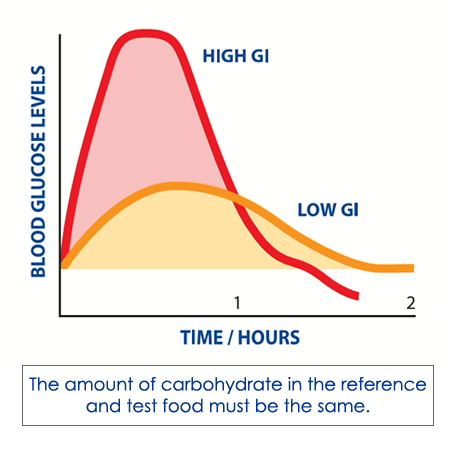
· Glycemic index (GI) & glycemic load (GL): Glycemic index (GI) is a system of assigning a number to carbohydrate-containing foods according to how much each food increases blood sugar. ‘Glycemic index diet’ usually refers to a specific diet plan that uses the index as the primary or only guides for meal planning. Many popular commercial diets, diet books, and diet websites are based on the glycemic index. The purpose of a glycemic index (GI) diet is to eat carbohydrate-containing foods that are less likely to cause large increases in blood sugar levels. The GI principle was first developed as a strategy for guiding food choices for people with diabetes. The glycemic index (GI) is a ranking of carbohydrates on a scale from 0 – 100 according to the extent to which they raise blood sugar levels after eating. Foods with a high GI are those which are rapidly digested and absorbed and result in marked fluctuations in blood sugar levels. Low-GI foods, by virtue of their slow digestion and absorption, produce gradual rises in blood sugar and insulin levels and have proven benefits for health.
· Low GI diets have been shown to improve both glucose and lipid levels in people with diabetes (type 1 and type 2).
· They have benefits for weight control because they help control appetite and delay hunger.
· Low GI diets also reduce insulin levels and insulin resistance.
· Recent studies from Harvard School of Public Health indicate that the risks of diseases such as type 2 diabetes and coronary heart disease (CHD) are strongly related to the GI of the overall diet.
· In 1999, the World Health Organisation (WHO) and Food and Agriculture Organisation (FAO) recommended that people in industrialized countries base their diets on low-GI foods in order to prevent the most common diseases of affluence, such as coronary heart disease (CHD), diabetes mellitus (DM) and obesity.
· An international GI database is maintained by Sydney University Glycemic Index Research Services in Sydney, Australia. The database contains the results of studies conducted there and at other research facilities around the world. There are various research methods for assigning a GI value to food. In general, the number is based on how much a food item raises blood glucose levels in healthy research participants compared with how much pure glucose raises their blood glucose.
· GI values are generally divided into three categories:
· a) Low GI: 1 – 55.
· b) Medium GI: 56 – 69.
· c) High GI: >_ 70.
· For example, raw carrots have a GI value of 35. This means that if someone eats enough carrots to consume 1.8 ounces (50 grams) of digestible carbohydrates (sugars and starches), his/her blood glucose level after eating the carrots will be 35% of the blood glucose level after eating 1.8 ounces (50 grams) of pure glucose. Comparing these values can help guide healthier food choices.
· Limitations of GI values: one limitation of GI values is that they don't reflect the likely quantity someone would eat of a particular food e.g., watermelon has a GI value of 80, which would put it in the category of food to avoid. But watermelon has relatively few digestible carbohydrates in a typical serving. In other words, someone has to eat a lot of watermelons to consume the standard test level of 1.8 ounces (50 grams) of digestible carbohydrates. Thus, researchers have developed the idea of glycemic load (GL).
· Glycemic load (GL) is a numerical value that indicates the change in blood glucose levels when someone eats a typical serving of the food, e.g., a 4.2-ounce (120-gram) serving of watermelon has a GL value of 5, which would identify it as a healthy food choice. Sydney University's table of GI values also includes GL values. The values are generally grouped in the following manner:
· a) Low GL: 1 – 10.
· b) Medium GL: 11 – 19.
· c) High GL: >_ 20.
· Glycemic index (GI) & glycemic load (GL) calculator:
· Hypoglycemia:
· Symptoms: sweating, hunger, trembling, anxiety, confusion, blurred vision, tachycardia (but may not appear if on beta–blockers).
· Causes:
· A) On newborns:
· i) Transient neonatal hypoglycemia: prematurity, intrauterine growth retardation (IUGR), perinatal asphyxia; maternal hyperglycemia due to diabetes or iatrogenic glucose administration; sepsis; prolonged fasting (e.g., due to inadequate breast milk or condition interfering with feeding).
· ii) Congenital hypopituitarism.
· iii) Congenital hyperinsulinism, several types, both transient and persistent.
· iv) Inborn errors of carbohydrate metabolism e.g., glycogen storage disease.
· B) On young children:
· i) Prolonged fasting: diarrheal illness in young children, e.g., Rotavirus gastroenteritis.
· ii) Idiopathic ketotic hypoglycemia.
· iii) Isolated GH (growth hormone) deficiency; hypopituitarism.
· iv) Insulin excess: hyperinsulinism due to several congenital disorders of insulin secretion; insulin injected for type 1 diabetes; hyperinsulinism hyperammonemia syndrome (HIHA) due to glutamate dehydrogenase 1 gene (in severe cases can cause mental retardation and epilepsy).
· v) Gastric dumping syndrome (after gastrointestinal surgery).
· vi) Other congenital metabolic diseases; some of the common include maple syrup urine disease and other organic acidurias; type 1 glycogen storage disease; type III glycogen storage disease (can cause less severe hypoglycemia than type I); phosphoenolpyruvate carboxykinase deficiency (metabolic acidosis and severe hypoglycemia); disorders of fatty acid oxidation; medium-chain acyl-CoA dehydrogenase deficiency (MCAD); familial leucine sensitive hypoglycemia.
· vii) Accidental ingestions including pharmacy misfiles; sulfonylureas, propranolol, and others; ethanol (mouthwash, alcoholic beverages).
· C) On young adults:
· i) Insulin-induced hypoglycemia: insulin injected for type 1 diabetes; factitious insulin injection (Munchausen syndrome); insulin-secreting pancreatic tumor (insulinoma); reactive hypoglycemia and idiopathic postprandial syndrome.
· ii) Sepsis.
· iii) Addison’s disease.
· D) On older adults:
· i) Insulin-induced hypoglycemia: insulin injected for diabetes; factitious insulin injection (Munchausen syndrome); excessive effects of oral anti-diabetic medication, beta-blockers, or drug interactions; insulin-secreting neuroendocrine tumor of the pancreas (insulinoma); alcohol-induced hypoglycemia often linked with ketoacidosis; alimentary (rapid jejunal emptying with exaggerated insulin response) after gastrectomy (dumping syndrome) or after bowel bypass surgery or resection; reactive hypoglycemia and idiopathic postprandial syndrome.
· ii) Tumor hypoglycemia, Doege – Potter syndrome.
· iii) Acquired adrenal insufficiency.
· iv) Acquired hypopituitarism.
· v) Immunopathologic hypoglycemia.
· Overview of causes:
· Α) Reactive:
· i) Hormonal deficiencies: hypoadrenalism (cortisol), hypopituitarism (growth hormone) (in children), glucagons deficiency (rare), and epinephrine (very rare).
· ii) Critical illnesses: cardiac, hepatic, and renal diseases; sepsis with multiorgan failure.
· iii) Exercise (in patients with diabetes treated with diabetes medications).
· iv) Pregnancy.
· v) Renal glycosuria.
· vi) Ketotic hypoglycemia of childhood.
· vii) Starvation.
· viii) Artifact.
· ix) Congenital enzyme deficiencies: hereditary fructose intolerance, galactosemia, and leucine sensitivity of childhood.
· Β) Exogenous insulin:
· 1) Insulin-producing tumors of the pancreas. Islet cell adenoma or carcinoma (insulinoma) is uncommon and is most often diagnosed in adults. It may occur as an isolated abnormality or as a component of the MEN (multiple endocrine neoplasia) type I syndrome. Carcinomas account for only 10% of insulin-secreting islet cell tumors. Approximately 60% of patients with insulinoma are female. Insulinomas are uncommon in persons younger than 20 years and are rare in those younger than 5 years. The median age at diagnosis is about 50 years, except in patients with MEN syndrome, in which the median age is in the mid-20s.
· 2) Non – beta-cell tumors. Hypoglycemia may be caused by large non–insulin-secreting tumors, most commonly retroperitoneal or mediastinal malignant mesenchymal tumors. The tumor secretes abnormal insulin-like growth factor (large IGF-II).
· C) Factitious hypoglycemia or self-induced hypoglycemia (can be seen in healthcare workers or in relatives who care for diabetic family members at home).
· D) Drug-induced. Hypoglycemia may be caused by the following drugs: ethanol (including propranolol plus ethanol), haloperidol, pentamidine, quinine, salicylates, and sulfonamides, oral hypoglycemics, phenylbutazone, insulin, bishydroxycoumarin, p-aminobenzoic acid, propoxyphene, stanozolol, hypoglycin, carbamate insecticide, disopyramide, isoniazid, methanol, methotrexate, tricyclic antidepressants, cytotoxic agents, organophosphates, didanosine, chlorpromazine, fluoxetine, sertraline, fenfluramine, trimethoprim, 6-mercaptopurine, thiazide diuretics, thioglycolate, tremetol, ritodrine, disodium ethylenediaminetetraacetic acid (EDTA), clofibrate, angiotensin-converting enzyme (ACE) inhibitors, and lithium.
· E) Fasting hypoglycemia.
· i) Nesidioblastosis: a rare cause of fasting hypoglycemia in infants and an extremely rare cause in an adult. It is characterized by a diffuse budding of insulin-secreting cells from the pancreatic duct epithelium and pancreatic microadenomas of such cells.
· ii) Causes of fasting hypoglycemia usually diagnosed in infancy or childhood include: inherited liver enzyme deficiencies that restrict hepatic glucose release (deficiencies of glucose-6-phosphatase, fructose-1,6-diphosphatase, phosphorylase, pyruvate carboxylase, phosphoenolpyruvate carboxykinase, or glycogen synthetase). Inherited defects in fatty acid oxidation, including that resulting from systemic carnitine deficiency and inherited defects in ketogenesis (3-hydroxy-3-methylglutaryl-CoA lyase deficiency) also may cause fasting hypoglycemia.
· Pseudohypoglycemia (artifactual hypoglycemia) - causes:
· a) Decreased capillary flow resulting in decreased glucose transport through the tissues and increased tissue extraction of glucose: Raynaud phenomena, acrocyanosis, peripheral vascular disease, Eisenmenger syndrome, and circulatory shock.
· b) Increased glycolysis by the leucocytes and red blood cells when there is a delay in interpreting the blood sample or separating plasma from the blood sample. Even in patients with normal leucocyte counts have shown an artificial decrease in glucose level (0.17 mmol/L/h) when it is allowed to clot at room temperature. Serum glucose concentration starts falling precipitously within 2 hours of collection if not refrigerated. A 90% lowering of glucose levels occurred when the blood was kept at room temperature for 2 hours.
· Pseudohypoglycemia may occur with: leukemia; leukemoid reaction (including eosinophilic leukemoid reaction due to underlying poorly differentiated carcinoma and hematopoietic cytokines-stimulated leukocytosis); polycythemia vera; blood samples containing high levels of other cell types due to the above mechanism as in chronic hemolytic anemia. Also may occur in primary red cell disorders associated with decreased survival and reticulocytosis.
· Moreover, African trypanosomiasis causes in vitro use of glucose.
· c) Hyperviscosity syndromes such as Waldenstrom macroglobulinemia and monoclonal gammaglobulinemia of undetermined significance (MGUS).
· d) Drugs such as ascorbic acid (especially high doses used in cancer therapy), dopamine, acetaminophen (paracetamol), and mannitol.
· Diagnostic algorithm for hypoglycemia:
· Uric acid: it is the final breakdown product of purine (*) catabolism. The liver and intestinal mucosa produce most of the uric acid. The kidneys eliminate 2/3 of the uric acid, with the GI (gastrointestinal) tract excretes the other one-third. Uric is a weak acid. The ionized forms of uric acid, urates, are present in synovial fluid and in plasma; approximately 98% exists as monosodium urate, with a pH of 7.4.
· The reference ranges for uric acid are:
· a) Men: 2.5-8 mg/dL.
· b) Women: 1.9–7.5 mg/dL.
· Serum urate concentrations in most children range from 3 – 4 mg/dL. During male puberty, levels begin to rise. Female levels remain low until menopause.
· Adult men have mean serum urate values of 6.8 mg/dL, and premenopausal women have mean serum urate values of 6 mg/dL.
· Values for women increase after menopause and approximate those of men. During adulthood, concentrations rise steadily and can vary with height, blood pressure, body weight, renal function, and alcohol intake.
· Causes of elevated uric acid levels include gout, kidney failure, destruction of massive amounts of nucleoproteins (leukemia, anemia, chemotherapy, toxemia of pregnancy, psoriasis, sickle cell anemia, hemolytic anemia, polycythemia, resulting pneumonia), drugs (especially diuretics and barbiturates, low-dose salicylates), lactic acidosis, hypothyroidism, chronic kidney disease, parathyroid diseases, metabolic acidosis, diet (high-protein weight-reducing diet, alcohol, liver, and sweet bread), chronic lead poisoning, Down syndrome, polycystic kidney disease, sarcoidosis, Lesch-Nyhan syndrome (**), von Gierke disease, and chronic berylliosis.
· Causes of decreased uric acid levels include drugs [including uricosuric drugs (salicylates, probenecid, allopurinol), estrogen, phenothiazines, indomethacin, corticotropin]; syndrome of inappropriate antidiuretic hormone secretion (SIADH) with hyponatremia; Wilson disease (***); Fanconi syndrome; acromegaly; celiac disease; and xanthinuria.
· (*) Purine is a heterocyclic aromatic organic compound. It consists of a pyrimidine ring fused to an imidazole ring. There are many naturally occurring purines. Two of the five bases in nucleic acids, adenine & guanine, are purines. In DNA these bases form hydrogen bonds with their complementary pyrimidines thymine & cytosine, respectively (this is called complementary base pairing). In RNA, the complement of adenine is uracil, instead of thymine. Other notable purines are hypoxanthine; xanthine; uric acid; and isoguanine.
· (**) Lesch Nyhan syndrome is a rare inherited disorder caused by a deficiency of the enzyme hypoxanthine-guanine phosphoribosyltransferase (HGPRT), produced by mutations in the HPRT gene on the X chromosome. It affects about one in 380,000 live births. The HGPRT deficiency causes a build-up of uric acid in all body fluids, resulting in both hyperuricemia and hyperuricosuria, associated with severe gout and kidney problems. Neurological signs include poor muscle control and moderate intellectual disability. These complications usually appear in the first year of life. Beginning in the second year of life, a particularly striking feature of LNS is self–mutating behaviors, characterized by lip and finger biting. Neurological symptoms include facial grimacing, involuntary writhing, and repetitive movements of the arms and legs. Also, some boys may develop megaloblastic anemia. This syndrome is present at birth in boys, however, there are a few rare cases in the world of affected females. Most, but not all, persons with this deficiency have severe mental and physical problems throughout life.
· (***) Wilson disease or hepatolenticular degeneration is an autosomal recessive genetic disorder in which copper (Cu) accumulates in tissues. It manifests as neurological or psychiatric symptoms and liver disease that may lead to liver failure and the need for liver transplantation. The condition is due to mutations in the Wilson disease protein (ATP7B) gene. Symptoms usually appear between the ages of 6 and 20 years, but cases in much older people have been described. Wilson's disease occurs in 1 to 4 per 100 000 people. Kayser – Fleischer rings are a pathognomic sign; these rings may be visible in the cornea of the eyes on slit-lamp examination as deposits of copper in a ring around the cornea.
· Gout & uric acid levels:
· a) Serum uric acid: the presence of hyperuricemia in the absence of symptoms is not diagnostic of gout. In addition, as many as 15% of patients with symptoms from gout may have normal serum uric acid levels at the time of their attack. Thus, the diagnosis of gout can be missed if the joint is not aspirated. Also, situations that decrease uric acid levels can trigger attacks of gout. In such cases, the patient’s medical records may reveal prior elevations of uric acid. Approximately 25% of the population has a history of elevated serum uric acid, but only a minority of patients with hyperuricemia develop gout. Thus, an abnormally high serum uric acid level does not indicate or predict gout. Gout is diagnosed by the presence of urate crystals in the synovial fluid or soft tissues. Also, some patients who present with a hot swollen joint and an elevated serum uric acid level, in fact, have infectious arthritis, which may be mismanaged if their synovial fluid is not examined. Asymptomatic hyperuricemia generally should not be treated. However, patients with levels higher than 11 mg/dL and over-excretion of uric acid are at increased risk for renal stones and renal impairment; therefore, renal function should be monitored in these individuals.
· The level of serum uric acid does correlate with the risk of developing gout. The 5-year risk for developing gout is approximately 0.6% if the level is below 7.9 mg/dL, 1% if it is 8 – 8.9 mg/dL, and 22% if it is higher than 9 mg/dL.
· b) Urinary uric acid: a 24-hour urinary uric acid evaluation is generally performed if uricosuric therapy is being considered. If patients excrete more than 800 mg of uric acid in 24 hours while eating a regular diet, they are overexpressors and thus overproducers of uric acid. These patients (approximately 10% of patients with gout) require the drug allopurinol instead of the medicine probenecid to reduce uric acid levels. Moreover, patients who excrete more than 1100 mg in 24 hours should undergo close renal function monitoring because of the risk of stones and urate nephropathy.
· Note: in patients in whom probenecid is contraindicated (e.g., those with a history of renal stones or renal insufficiency), a 24-hour urine test of uric acid excretion need not be performed, because the patient clearly will need allopurinol.
· Uric acid in diet and recommendations for people with gout:
· Uric acid in the diet: In humans, purines are excreted as uric acid. Purines are found in high amounts in animal food products, such as liver and sardines. A moderate amount of purine is also contained in beef, pork, poultry, fish and seafood, asparagus, cauliflower, spinach, mushrooms, green peas, lentils, dried peas, beans, oatmeal, wheat bran, and wheat germ.
· Milk products reduce the risk of gout, whereas total protein intake has no effect.
· Recommendations for specific foods or supplements for people with gout (according to Mayo clinic): a) High-purine vegetables. Studies have shown that vegetables high in purines do not increase the risk of gout or recurring gout attacks. A healthy diet based on lots of fruits and vegetables can include high-purine vegetables, such as asparagus, spinach, peas, cauliflower, or mushrooms. Someone with gout can also eat beans or lentils, which are moderately high in purines but are also a good source of protein. b) Organ and glandular meats. People with gout should avoid meats such as liver, kidney, and sweetbreads, which have high purine levels and contribute to high blood levels of uric acid. c) Selected seafood. People with gout should avoid the following types of seafood, which are higher in purines than others: anchovies, herring, sardines, mussels, scallops, trout, haddock, mackerel, and tuna. d) Alcohol. The metabolism of alcohol in the body is thought to increase uric acid production, and alcohol contributes to dehydration. Beer is associated with an increased risk of gout and recurring attacks, as are distilled liquors to some extent. The effect of wine is not as well-understood. e) Vitamin C. Vitamin C may help lower uric acid levels. f) Coffee. Some research suggests that moderate coffee consumption may be associated with a reduced risk of gout, particularly with regular caffeinated coffee. Drinking coffee may not be appropriate for other medical conditions. g) Cherries. There is some evidence that eating cherries is associated with a reduced risk of gout attacks (Reference (Retrieved: December 22, 2015): http://www.mayoclinic.org/healthy-lifestyle/nutrition-and-healthy-eating/in-depth/gout-diet/art-20048524 ).
· The results of a study on a nationally representative sample of adults in the US suggest that higher levels of meat and seafood consumption are associated with higher serum levels of uric acid but that total protein intake is not. Dairy consumption was inversely associated with the serum uric acid level (Reference: http://www.ncbi.nlm.nih.gov/pubmed/15641075 ).
No comments:
Post a Comment